By B.G Ramaprasad
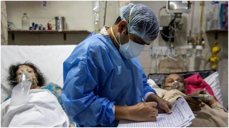
Cases of mucormycosis, or “black fungus,” a potentially serious condition that causes blurred or double vision, chest pain, and breathing difficulties, have surged in India, mostly among COVID-19 patients. At least 8,848 such cases have been found across the country as of May 21, according to the government. The disease is caused by fungal spores found in soil and organic matter, usually inhaled by humans from the air. The mould enters the body and then manifests around the nose and eye sockets, causing the nose to blacken, and if not stopped will move fatally into the brain.
Black fungus is caused by organisms called mucormycetes, which can enter the body through breathing or skin injuries. These are naturally present in soil and decaying organic matter, but once inside humans, they can infect air pockets behind the forehead, nose, cheekbones and between the eyes and teeth.
Mucormycosis, previously known as zygomycosis (and especially for rhino-orbital-cerebral mucormycosis, sometimes called black fungus) is a serious fungal infection, generally in people with less ability to fight infection. Symptoms depend on the part of the body infected. It most commonly infects the sinuses and brain resulting in a runny nose, one-sided facial swelling and pain, headache, fever, and tissue death. Other forms of disease may infect the lungs, stomach and intestines, and skin. It is generally spread by breathing in, eating food contaminated by, or getting spores of molds of the Mucorales type in an open wound. These fungi are frequently present in the air, in decomposing organic matter such as rotting fruit and vegetables, leaves, and animal manure, but do not usually affect people. It is not transmitted between people. Risk factors include diabetes (particularly DKA), cancer, organ transplant, iron overload, long-term steroids or immunosuppressant use, and to a lesser extent in HIV/AIDS. Diagnosis is by biopsy and culture, with medical imaging to help determine the extent of disease. It may appear similar to aspergillosis.
Signs and symptoms

- Signs and symptoms of Mucormycosis relate to where the infection is. Infection usually begins in the mouth or nose and enters the central nervous system via the eyes.
- Fever, cough, chest pain, and difficulty breathing, or coughing up blood, can occur when the lungs are involved.[6] A tummy ache, nausea, vomiting, and bleeding can occur when the gastrointestinal tract is involved. Affected skin may appear as a dusky reddish tender patch with a darkening center due to tissue death. There may be an ulcer and it can be very painful.
- Invasion into the blood vessels can result in thrombosis and subsequent death of surrounding tissue due to a loss of blood supply.
Causes :
Mucormycosis is a fungal infection caused by fungi in the order Mucorales.In most cases it is due to an invasion of the genera Rhizopus and Mucor, common bread molds. Most fatal infections are caused by Rhizopus oryzae. It is less likely due to Lichtheimia, and rarely due to Apophysomyces. Others include Cunninghamella, Mortierella, and Saksenaea.The fungal spores are in indoor and outdoor air, in dust, can be found on for instance moldy bread and fruit and are breathed in frequently, but cause disease only in some people. In addition to being breathed in to be deposited in the nose, sinuses and lungs, the spores can also enter the skin through a cut or open wound, or grow in the intestine if eaten.Once deposited, the fungus grows branch-like filaments which invade blood vessels, causing clots to form and surrounding tissues to die. Other reported causes include contaminated
wound dressings.
Risk factors :
Predisposing factors for mucormycosis include conditions where people are less able to fight infection, have a low neutrophil count or metabolic acidosis. Risk factors include poorly controlled diabetes mellitus (particularly DKA), organ transplant, iron overload, cancers such as lymphomas, kidney failure, long-term corticosteroid and immunosuppressive therapy, liver disease and severe malnutrition. Other risk factors include tuberculosis.
Mechanism :
Most people are frequently exposed to Mucorales without developing the disease. Mucormycosis is generally spread by breathing in, eating food contaminated by, or getting spores of molds of the Mucorales type in an open wound. It is not transmitted between people.
Diagnosis :
Diagnosis requires identifying the mold in the affected tissue by biopsy and confirming it with a fungal culture. Because the causative fungi occur all around, a culture alone is not decisive. Tests may also include culture and direct detection of the fungus in lung fluid, blood, serum, plasma and urine.
Imaging :
Imaging is often performed, such as CT scan of lungs and sinuses.[38] Signs on chest CT scans, such as nodules, cavities, halo signs, pleural effusion and wedge-shaped shadows, showing invasion of blood vessels may suggest a fungal infection, but does not confirm mucormycosis.
CT scan images of mucormycosis can be useful to distinguish mucormycosis of the orbit and cellulitis of the orbit, but imaging may look identical to those of aspergillosis.
Prevention :
Preventive measures include wearing a face mask in dusty areas, avoiding direct contact with water-damaged buildings, and protecting skin, feet and hands where there is exposure to soil or manure such as gardening or certain outdoor work. In high-risk groups such as organ transplant, antifungal drugs may be given as a preventative.
Treatment :
If mucormycosis is suspected, Amphotericin B is initially given slowly into a vein, then given daily for the next
14 days. Amphotericin B is sometimes continued for longer.In 2015, without a randomised control trial,
The FDA approved Isavuconazole as a treatment for mucormycosis. Posaconazole is an alternative. Surgical removal of the “fungus ball” is then indicated. . Surgery can be very drastic, and in some cases of disease involving the nasal cavity and the removal of infected brain tissue may be required. Removal of the palate, nasal cavity, or eye structures
can be very disfiguring. Sometimes more than one operation is required.
Naming :
Arnold Paltauf coined the term “Mycosis Mucorina” in 1885, after describing a case with systemic symptoms involving the sinus, brain and gastrointestinal tract, following which the term “mucormycosis” became popular. “
Notable outbreaks :
The disease has been reported in natural disasters and catastrophes; 2004 Indian Ocean tsunami and the 2011 Missouri tornado.[20][49] The first international congress on mucormycosis was held in Chicago in 2010, set up by the Hank Schueler 41 & 9 Foundation, which was established in 2008 for the research of children with leukaemia and fungal infections. A cluster of infections occurred in the wake of the 2011 Joplin tornado. By July 19, 2011 a total of 18 suspected cases of mucormycosis of the skin had been identified, of which 13 were confirmed.
COVID-19–associated mucormycosis :

Countries where COVID-associated mucormycosis has been detected as of June 2021COVID-19-associated mucormycosis, commonly referred to as black fungus, is the association of rhinocerebral mucormycosis with COVID-19. It has been reported around the nose, eyes and brain – a clinical manifestation sometimes referred to as “rhino-orbital-cerebral (ROC) mucormycosis”. The condition does not spread person to person and is not contagious.
Reports of COVID-associated mucormycosis have generally been rare. A review of the medical literature traced eight cases reported around the world by 9 January 2021. In these reports, the most common risk factor for mucormycosis was diabetes. Most cases presented during hospitalization (often 10–14 days after admission), and all but one of the affected people died. Early aggressive treatment is considered essential. (It has been estimated that between 40% and 80% . People who contract any form of mucormycosis die from the disease, depending on the site of infection and underlying health conditions.
_cases.png)